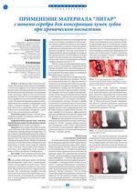
Применение материала “литар” с ионами серебра для консервации лунок зубов при хроническом воспалении


Научно-практический журнал Институт Стоматологии №1(94), апрель 2022
стр. 76-77
Аннотация
Атрофия костной ткани после удаления зуба вызывает убыль объема альвеолярного отростка и дефицит кости при последующей имплантации. Для предотвращения данных нежелательных явлений проводится консервация лунок удаленных зубов костно-пластическим материалом “ЛитАр”. В состав материала входят ионы серебра для антисептической обработки тканей. 30 пациентам проведено удаление зубов с хроническим воспалением и консервация лунок зубов с помощью материала “ЛитАр” с серебром. Отмечается сохранение объема костной ткани после имплантации материала, а также отсутствие воспалительных явлений.
Аннотация (англ)
Bone atrophy after tooth extraction causes a decrease in the volume of the alveolar bone and deficiency during subsequent implantation. To prevent these undesirable phenomena, the sockets of the extracted teeth are preserved with the bone-plastic material “Litar”. The material contains silver ions for antiseptic processing of tissues. 30 patients underwent extraction of teeth with chronic inflammation and preservation of tooth sockets using the material “Litar” with silver. Preservation of the volume of bone tissue after implantation of the material is noted, as well as the absence of inflammation.
Ключевые Слова
материал “ЛитАр”, наноразмерное серебро, консервация лунки зуба, атрофия костной ткани.
Ключевые Слова (англ)
“LitAr” material, nanoscale silver, preservation of the tooth socket, bone atrophy.
Список литературы
ЛИТЕРАТУРА / REFERENCES:
1. Конев В.А., Божкова С.А., Трушников В.В. [и др.]. Динамика тканевых изменений при одно- и двухэтапном лечении хронического остеомиелита с использованием биорезорбируемого материала, импрегнированного ванкомицином (сравнительное экспериментально-морфологическое исследование) // Гены и Клетки. - 2021. - Т. 16. - № 1. - С. 29-36. [Konev V.A., Bozhkova S.A., Trushnikov V.V. [i dr.]. Dinamika tkanevyh izmenenij pri odno- i dvuhetapnom lechenii hronicheskogo osteomielita s ispol’zovaniem biorezorbiruemogo materiala, impregnirovannogo vankomicinom (sravnitel’noe eksperimental’no-morfologicheskoe issledovanie) // Geny i Kletki. - 2021. - T. 16. - № 1. - S. 29-36.]
2. Литвинов С.Д., Лепилин И.Н. Применение материала “ЛитАр” на альгинатной основе для консервации лунки зуба // Вестник медицинского института “РЕАВИЗ”: реабилитация, врач и здоровье. - 2017. - № 6 (30). - С. 158-165. [Litvinov S.D., Lepilin I.N. Primenenie materiala “LitAr” na al’ginatnoj osnove dlya konservacii lunki zuba // Vestnik medicinskogo instituta “REAVIZ”: reabilitaciya, vrach i zdorov’e. - 2017. - № 6 (30). - S. 158-165.]
3. Al-Moraissi E.A., Alkhutari A.S., Abotaleb B., Altairi N.H., Fabbro M.D. Do osteoconductive bone substitutes result in similar bone regeneration for maxillary sinus augmentation when compared to osteogenic and osteoinductive bone grafts? A systematic review and frequentist network meta-analysis. Int. J. Oral. Maxillofac. Surg. 2019;19:31163-31164.
4. Feng Q.L., Wu J., Chen G.Q., Cui F.Z., Kim T.N., Kim J.O. A mechanistic study of the antibacterial effect of silver ions on Escherichia coli and Staphylococcus aureus. J. Biomed Mater Res 2000;52:662-8.
5. Franci G., Falanga A., Galdiero S., Palomba L., Rai M., Morelli G., Galdiero M. Silver nanoparticles aspotential antibacterial agents.Molecules2015,20, 8856-8874.
6. Kędziora A., Speruda M., Krzyżewska E., Rybka J., Łukowiak A., & Bugla-Płoskońska G. (2018). Similarities and Differences between Silver Ions and Silver in Nanoforms as Antibacterial Agents. International Journal of Molecular Sciences, 19(2), 444.
7. Mastrangelo F., Quaresima R., Grilli A., Tettamanti L., Vinci R., Sammartino G., Tetè S., Gherlone E. A comparison of bovine bone and hydroxyapatite scaffolds during initial bone regeneration: An in vitro evaluation. Implant Dent. 2013;22:613-622.
8. Orgeas G.V., Clementini M., Risi V.D., Sanctis M.D. Surgical Techniques for Alveolar Socket Preservation: A Systematic Review. Int. J. Oral Maxillofac. Implants. 2013;28:1049-1061.
9. Zhang K., Melo M.A., Cheng L., Weir M.D., Bai Y., Xu H.H. Effect of quaternary ammonium and silver nanoparticle-containing adhesives on dentin bond strength hand dental plaque microcosm biofilms. Dent Mater 2012;28:842
Другие статьи из раздела «Клиническая стоматология»
- Комментарии
Загрузка комментариев...
|
Поделиться:
|

 PDF)
PDF)


