Эндодонтическое лечение первого моляра нижней челюсти шестью корневыми каналами — два клинических случая и обзор литературы


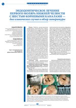
Наиболее распространенная конфигурация нижнечелюстного первого моляра предполагает наличие двух корней и трех корневых каналов. Цель данной работы состоит в демонстрации двух редких случаев анатомической конфигурации с шестью корневыми каналами в первых левых молярах нижней челюсти, диагностированными в процессе эндодонтической терапии. Лечение корневых каналов проводилось с применением дентального операционного микроскопа. Ультразвуковое исследование сообщений между мезиальными и дистальными корневыми каналами показало наличие промежуточных каналов. Исследования больших выборочных групп населения и систематические обзорные исследования не выявили ни одного случая конфигурации с шестью корневыми каналами в первых нижнечелюстных молярах. Можно обнаружить три разных вида конфигурации пульповой камеры. Могут быть в наличии два или три корня, при этом чаще встречается конфигурация мезиального корня Типа 8 и дистального корня Типа 12. Также рассматриваются некоторые идеи в связи с необходимыми в таких случаях техническими подходами.
Endodontic therapy, Molar, Root canal anatomy, Root canal preparation
1. Wolcott J., Ishley D., Kennedy W., Johnson S., Minnich S., Meyers J. A 5 yr clinical investigation of second mesiobuccal canals in endodontically treated and retreated maxillary molars. J. Endod. 2005; 31(4): 262-64.
2. de Pablo O., Estevez R., Sanchez M., Heilborn C., Cohenca N. Root anatomy and canal confiuration of the permanent mandibular fist molar: a systematic review. J. Endod. 2010; 36(12): 1919-31.
3. Vertucci F.J. Root canal anatomy of the human permanent teeth. Oral Surg. 1984; 58(5): 589-99.
4. Gulabivala K., Aung T.H., Alavi A., Ng Y.L. Root and canal morphology of Burmese mandibular molars. Int. Endod J. 2011; 34(5): 359-70.
5. Caliskan M., Pehlivan Y., Sepetcioglu Figen S., Turkun M., Tuncer S. Root canal morphology of human permanent teeth in a Turkish population. J. Endod. 1995; 21(4): 200-04.
6. Martinez-Berna A., Badanelli P. Mandibular fist molars with six root canals. J. Endod. 1985; 11(8): 348-52.
7. Reeh E.S. Seven canals in lower fist molar. J. Endod. 1998; 24(7): 497-99.
8. Ghoddusi J., Naghavi N., Zarei M., Rohani E. Mandibular fist molar with four distal canals. J. Endod. 2007; 33(12): 1481-83.
9. Aminsobhani M., Shokouhinejad N., Ghabraei S., Bolhari B., Ghorbanzadeh A. Retreatment of a 6-canalled mandibular fist molar with four mesial canals: a case report. Iran Endod J. 2010; 5(3): 138-40.
10. Ryan J., Bowles W., Baisden M., McClanahan S. Mandibular fist molar with six separated canals. J. Endod. 2011; 37(6): 878-80.
11. Gupta S., Jaiswal S., Arora R. Endodontic management of permanent mandibular left fist molar with six root canals. Contemp Clin Dent. 2012; 3(Suppl 1): S130-S133. doi: 10.4103/0976-237X.95124.
12. Baziar H., Daneshvar F., Mohammadi A., Jafarzadeh H. Endodontic management of a mandibular fist molar with four canals in a distal root by using cone-beam computed tomography: a case report. J. Oral Maxillofac Res. 2014; 5(1): e5.
13. Hasan M., Rahman M., Saad N. Mandibular fist molar with six root canals: a rare entity. BMJ Case Rep. 2014; 2014:bcr2014205253. doi: 10.1136/bcr-2014-205253.
14. Sinha N., Singh B., Langaliya A., Mirdha N., Huda I., Jain A. Cone beam computed topographic evaluation and endodontic management of a rare mandibular fist molar with four distal canals. Case Rep Dent. 2014;2014:306943. doi:10.1155/2014/306943. Epub 2014 Dec 1.
15. Patel S., Durack C., Abella F., Roig M., Shemesh H., Lambrechts P., et al. European Society of Endodontology position statement: The use of CBCT in Endodontics. Int Endod J. 2014; 47(6): 502-04.
16. Martins J.N. Endodontic treatment of a maxillary fist molar with seven root canals confimed with cone beam computer tomography - case report. J Clin Diagn Res. 2014; 8(6): 13-15.
17. Macedo R.G., Wesselink P.R., Zaccheo F., Fanali D., van der Sluis LWM. Reaction rate of NaOCl in contact with bovine dentine: effect of activation, exposure time, concentration and pH. Int. Endod J. 2010; 43(12): 1108-15.
Другие статьи из раздела «Клиническая стоматология»
- Комментарии
|
Поделиться:
|

 PDF)
PDF)


