
Биологические факторы и свойства имплантатов, обеспечивающие супракрестальную защиту — Двойной Биологический Барьер

Научно-практический журнал Институт Стоматологии №1 (110), апрель 2026
стр. 89-93
Стоимость:
250 руб.
(в формате  PDF)
PDF)
PDF)| В корзину |
Ссылка на elibrary.ru
Аннотация
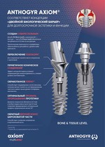
В статье представлена клиническая концепция Двойного Биологического Барьера (Double Biological Barrier, DBB), обеспечивающая долгосрочную стабильность тканей в зоне соединения имплантата с абатментом. Обзор объединяет знания о регенерации костной и мягких тканей с конструктивными особенностями имплантационных систем: шероховатой обработкой плеча имплантата, эффектом переключения платформ, субкрестальным позиционированием дентального имплантата, коническим соединением абатмента, формой и высотой трансгингивальных компонентов, а также контролируемым профилированием костного гребня и оптимальным объёмом кератинизированной десны. Формирование костного и мягкотканного барьеров создаёт механическую и биологическую защиту периимплантатных структур, снижает риск воспалительных осложнений и способствует стабилизации маргинальной кости и мягкотканного контура. Концепция Двойной Биологический Барьер (ДББ) представляет собой клинически обоснованный подход к планированию и реализации дентальной имплантации, направленный на повышение предсказуемости долгосрочного результата лечения.
Аннотация (англ)
This article presents the Double Biological Barrier (DBB) concept, ensuring long-term stability of tissues at the implant—abutment interface. The model integrates knowledge of bone and soft tissue regeneration with implant design features, including roughened collar surfaces, platform switching, subcrestal placement, conical abutment connection, transgingival component design, controlled bone ridge profiling, and optimal keratinized mucosa volume. Formation of bone and soft tissue barriers provides mechanical and biological protection of peri-implant structures, reduces the risk of inflammatory complications, and supports marginal bone and soft tissue contour stability. DBB represents a clinically justified approach to implant treatment planning and implementation, enhancing predictability of long-term outcomes.
Ключевые Слова
дентальные имплантаты; остеоинтеграция; периимплантит; маргинальная резорбция кости; интерфейс «имплантат—абатмент»; периимплантатные мягкие ткани; биологическая ширина; двойной биологический барьер.
Ключевые Слова (англ)
dental implants; osseointegration; peri-implantitis; marginal bone loss; implant—abutment interface; peri-implant soft tissue; biological width; double biological barrier.
Список литературы
1. Adel K.M., Abd Elfatah S., Abd El-Mageed Katamish M. Crestal bone loss of standard implant versus platform switch implant design using minimal invasive technique // J. Fac Dent J. 2016;7:16-21. doi:10.1016/j.fdj.2016.09.001.
2. Anitua E., Alkhraisat M.H., Eguia A. On peri-implant bone loss theories: trying to piece together the jigsaw. Cureus. 2023;15:e33237. doi:10.7759/cureus.33237.
3. Comuzzi L., Ceddia M., Di Pietro N., Inchingolo F., Inchingolo A.M., Romasco T. et al. Crestal and subcrestal placement of Morse cone implant-abutment connection implants: an in vitro finite element analysis study // Biomedicines. - 2023;11:3077. doi:10.3390/biomedicines11113077.
4. Fickl S., Zuhr O., Stein J.M., Hürzeler M.B. Peri-implant bone level around implants with platform-switched abutments // Int J. Oral Maxillofac Implants. - 2010;25:577-581.
5. Gualini F., Salina S., Rigotti F., Mazzarini C., Longhin D., Grigoletto M. et al. Subcrestal placement of dental implants with an internal conical connection of 0.5 mm versus 1.5 mm // Eur J. Oral Implantol. 2017;10:73-82.
6. Kikuchi T., Wada M., Mameno T., Hasegawa D., Serino G., Ikebe K. Longitudinal study on the effect of keratinized mucosal augmentation // Peer J. 2022;10:e13598.
7. Koodaryan R., Hafezeqoran A. Evaluation of implant collar surfaces for marginal bone loss. Biomed Res Int. 2016;2016:4987526.
8. Koutouzis T., Gholami F., Reynolds J., Lundgren T., Kotsakis G.A. Abutment disconnection/reconnection affects peri-implant marginal bone levels // Int J. Oral Maxillofac Implants. - 2017;32:575-581.
9. Lin H.K., Lin J.C.Y. et al. Peri-implant marginal bone changes around implants with platform switching // J. Pers Med. - 2022;12:1226.
10. Messias A., Nicolau P., Guerra F. Titanium dental implants with different collar design // Clin Oral Implants Res. - 2019;30:20-48.
11. Monje A., Roccuzzo A., Buser D., Wang H.L. Influence of buccal bone wall thickness // Clin Oral Implants Res. 2023;34:8-27.
12. Palacios-Garzón N., Velasco-Ortega E., López-López J. Bone loss in implants placed at subcrestal and crestal level. Materials. - 2019;12:154.
13. Pellegrini G., Canullo L., Dellavia C. Histological features of peri-implant bone subjected to overload. Ann Anat. - 2016;206:57-63.
14. Quispe López N., Guadilla Y., Gómez Polo C., López Valverde N., Flores Fraile J., Montero J. Influence of implant depth and abutment height // J. Dent. - 2024;148:105264.
15. Ramanauskaite A., Schwarz F., Sader R. Influence of width of keratinized tissue // Clin Oral Implants Res. 2022;33:8-31.
16. Riberti N. et al. Case report of a dental implant with conometric abutment. Clin Pract. 2024;14:556-569.
17. Siqueira R.A.C., Gonçalves Junior R.S., Santos P.G.F., Sartori I.A.M., Wang H.L., Fontão F.N.G.K. Effect of implant placement depth // Clin Oral Implants Res. - 2020;31:282-293.
18. Soulami S., Slot D.E., van der Weijden F. Implant-abutment emergence angle and profile // Clin Exp Dent Res. - 2022;8:795-806.
19. Sun Y., Yang J., Chen K., Li Z., Chen Z., Huang B. Crestal vs subcrestal placement of implants // Clin Implant Dent Relat Res. - 2023;25:948-959.
20. Veljanovski D., Atanasovska-Stojanovska A., Pivkova-Veljanovska A., Mijiritsky E., Bollen C. Soft tissue thickness and implant placement. Open Access Maced J. Med Sci. 2021;9:257-263.
21. Zipprich H., Weigl P., Ratka C., Lange B., Lauer H.C. Micromovements at the implant-abutment interface: Measurement, causes, and consequences. Implantologie. - 2007;15(1):31-46.
22. Yang F., Ruan Y., Liu Y., Chen J., Chen Y., Zhang W. et al. Abutment mechanical complications of Morse taper connection. Clin Implant Dent Relat Res. 2022;24:683-695.
2. Anitua E., Alkhraisat M.H., Eguia A. On peri-implant bone loss theories: trying to piece together the jigsaw. Cureus. 2023;15:e33237. doi:10.7759/cureus.33237.
3. Comuzzi L., Ceddia M., Di Pietro N., Inchingolo F., Inchingolo A.M., Romasco T. et al. Crestal and subcrestal placement of Morse cone implant-abutment connection implants: an in vitro finite element analysis study // Biomedicines. - 2023;11:3077. doi:10.3390/biomedicines11113077.
4. Fickl S., Zuhr O., Stein J.M., Hürzeler M.B. Peri-implant bone level around implants with platform-switched abutments // Int J. Oral Maxillofac Implants. - 2010;25:577-581.
5. Gualini F., Salina S., Rigotti F., Mazzarini C., Longhin D., Grigoletto M. et al. Subcrestal placement of dental implants with an internal conical connection of 0.5 mm versus 1.5 mm // Eur J. Oral Implantol. 2017;10:73-82.
6. Kikuchi T., Wada M., Mameno T., Hasegawa D., Serino G., Ikebe K. Longitudinal study on the effect of keratinized mucosal augmentation // Peer J. 2022;10:e13598.
7. Koodaryan R., Hafezeqoran A. Evaluation of implant collar surfaces for marginal bone loss. Biomed Res Int. 2016;2016:4987526.
8. Koutouzis T., Gholami F., Reynolds J., Lundgren T., Kotsakis G.A. Abutment disconnection/reconnection affects peri-implant marginal bone levels // Int J. Oral Maxillofac Implants. - 2017;32:575-581.
9. Lin H.K., Lin J.C.Y. et al. Peri-implant marginal bone changes around implants with platform switching // J. Pers Med. - 2022;12:1226.
10. Messias A., Nicolau P., Guerra F. Titanium dental implants with different collar design // Clin Oral Implants Res. - 2019;30:20-48.
11. Monje A., Roccuzzo A., Buser D., Wang H.L. Influence of buccal bone wall thickness // Clin Oral Implants Res. 2023;34:8-27.
12. Palacios-Garzón N., Velasco-Ortega E., López-López J. Bone loss in implants placed at subcrestal and crestal level. Materials. - 2019;12:154.
13. Pellegrini G., Canullo L., Dellavia C. Histological features of peri-implant bone subjected to overload. Ann Anat. - 2016;206:57-63.
14. Quispe López N., Guadilla Y., Gómez Polo C., López Valverde N., Flores Fraile J., Montero J. Influence of implant depth and abutment height // J. Dent. - 2024;148:105264.
15. Ramanauskaite A., Schwarz F., Sader R. Influence of width of keratinized tissue // Clin Oral Implants Res. 2022;33:8-31.
16. Riberti N. et al. Case report of a dental implant with conometric abutment. Clin Pract. 2024;14:556-569.
17. Siqueira R.A.C., Gonçalves Junior R.S., Santos P.G.F., Sartori I.A.M., Wang H.L., Fontão F.N.G.K. Effect of implant placement depth // Clin Oral Implants Res. - 2020;31:282-293.
18. Soulami S., Slot D.E., van der Weijden F. Implant-abutment emergence angle and profile // Clin Exp Dent Res. - 2022;8:795-806.
19. Sun Y., Yang J., Chen K., Li Z., Chen Z., Huang B. Crestal vs subcrestal placement of implants // Clin Implant Dent Relat Res. - 2023;25:948-959.
20. Veljanovski D., Atanasovska-Stojanovska A., Pivkova-Veljanovska A., Mijiritsky E., Bollen C. Soft tissue thickness and implant placement. Open Access Maced J. Med Sci. 2021;9:257-263.
21. Zipprich H., Weigl P., Ratka C., Lange B., Lauer H.C. Micromovements at the implant-abutment interface: Measurement, causes, and consequences. Implantologie. - 2007;15(1):31-46.
22. Yang F., Ruan Y., Liu Y., Chen J., Chen Y., Zhang W. et al. Abutment mechanical complications of Morse taper connection. Clin Implant Dent Relat Res. 2022;24:683-695.
Другие статьи из раздела «Обзор литературы в стоматологии»
- Комментарии
Загрузка комментариев...
|
Поделиться:
|


