Лучевая диагностика рассекающего остеохондрита височно-нижнечелюстного сустава

Научно-практический журнал Институт Стоматологии №3 (60), сентябрь 2013
Стоимость:
Бесплатно
(в формате  PDF)
PDF)
PDF)Аннотация
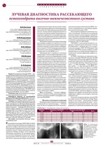
Рассекающий остеохондрит (РОХ) — редкое заболевание неизвестной этиологии, поражающее эпифизы костей. В литературе описаны единичные случаи поражения РОХ височно-нижнечелюстного сустава (ВНЧС). Цель исследования — изучение информативности линейной (ТМГ), компьютерной (КТ), магнитно-резонансной (МРТ) томографии в диагностике РОХ ВНЧС. Чувствительность и точность лучевой диагностики РОХ ВНЧС разные, что связано с местом поражения суставной поверхности, стадийным течением заболевания. ТМГ ВНЧС имеет более низкие значения чувствительности, точности выявления РОХ ВНЧС, чем КТ и МРТ.
Аннотация (англ)
Osteochondritis dissecans (OD) is a rare disease of unknown etiology, affecting the epiphyseal bones. In the literature are described isolated cases of the defeat of the OD the temporomandibular joint (TMJ). The purpose of research — studying of the informativeness of linear (TMG), computer (CT), magnetic resonance imaging (MRI) in the diagnosis of the OD TMJ. The sensitivity and accuracy of x-ray diagnostics of the OD TMJ different, that is connected with a place of destruction of articular surfaces, course of the disease. TMG TMJ has lower values of sensitivity, accuracy of detection of the OD TMJ than CT and MRI.
Ключевые Слова
рассекающий остеохондрит, височно-нижнечелюстной сустав
Ключевые Слова (англ)
osteochondritis dissecans, temporomandibular joint.
Список литературы
1. Райзер М., Бауер-Мельник А., Глассер К. Лучевая диагностика. Костно-мышечная система // Пер. с англ. под общ. ред. Н.Б.Петровой. - М.: МЕДпресс-информ, 2011. - 384 с.
2. Atik OS., Esen E., Tokgoz N. et al. Osteochondritis dissecans with subchondral bone cyst of the femoral condyle: a novel surgical technique of treatment // Eklem Hastalik Cerrahisi. - 2009. -Vol. 20. - № 3. - Р. 174-177.
3. Campos P.S., Freitas C.E., Pena N. et al. Osteochondritis dissecans temporomandibular joint // Dentomaxillofac Radiol. - 2005. - Vol.34. - № 3. - Р. 193-197.
4. Emre TY., Cift H., Seyhan B. et al. Midtern results of biologic fixation or mosaicplasty and drilling in osteochondritis dissecans // Indian J Orthop. - 2011. - Vol.45. - № 5. - Р. 445-449.
5. Jans LB., Ditchfield M., Anna G. et al. MR imaging findings and MR criteria for instability in osteochondritis dissecans of the elbow in chilbren // Eur J Radiol. - 2012. - Vol.81. - № 6. - Р. 1306-1310.
6. Laor T., Zbojniewicz AM., Eismann EA., Wall EJ. Juvenile osteochondritis dissecans: is it a growth disturbance of the secondary physis of the epiphysis? // Am J Roentgenol. - 2012. - Vol.199. - № 5. - Р. 1121-1128.
7. Orhan K., Arslan A., Kocyigit D.Temporomandibular joint osteochondritis dissecans: case report // Oral Surg Oral Med Oral Pathol Oral Radiol Endod. - 2006. - Vol.102. - № 4. - Р. 41-46.
8. Orr JD., Dawson LK., Garcia EJ., Kirk KL. Incidence of osteochondral lesions of the talus in the United States military // Foot Ankle Int. - 2011. - Vol.32. - № 10. - Р. 948-954.
9. Rammal H., Gicquel P., Schneider L. et al. Juvenile osteochondritis of femoral condyles: treatment with transchondral drilling. Analysis of 40 cases // J Child Orthop. - 2010. - Vol.4. - № 1. - Р. 39-44.
10. Samora WP., Chevillet J., Adler B. et al. Juvenile osteochondritis dissecans of the knee: predictors of lesion stability // J Pediatr Orthop. - 2012. - Vol.32. - № 1. - Р. 1-4.
Другие статьи из раздела «Клиническая стоматология»
- Комментарии
Загрузка комментариев...
|
Поделиться:
|


